Home
Uncategories
Anatomy Diagram Rib Area / Intercostal Muscles - Function, Area & Course - Human Anatomy | Kenhub - YouTube - The serratus anterior muscle originates from a roughened area near the middle of.
Anatomy Diagram Rib Area / Intercostal Muscles - Function, Area & Course - Human Anatomy | Kenhub - YouTube - The serratus anterior muscle originates from a roughened area near the middle of.
Anatomy Diagram Rib Area / Intercostal Muscles - Function, Area & Course - Human Anatomy | Kenhub - YouTube - The serratus anterior muscle originates from a roughened area near the middle of.. This free online diploma course introduces you to the basic anatomy and physiology of important systems in the body. 12 pairs of anatomical ribs are present in thoracic region — originating from 1st to 12th (t1 to t12) thoracic vertebrae. Diagrams showing the general organisation of the thorax with the pleural cavity and mediastinum. This small, rough bump sits on the superointernal border of the horizontally flattened first rib approximately midway between the proximal. These are large areas of the cerebral cortex that receive sensory input from multiple different sensory modalities and various association areas and help make associations between various kinds of sensory info.
They also have a role in. Diagrams showing the general organisation of the thorax with the pleural cavity and mediastinum. The ribs are a set of twelve paired bones which form the protective 'cage' of the thorax. Instant anatomy is a specialised web site for you to learn all about human anatomy of the body with diagrams, podcasts and revision questions. Posterior, anterior and limbic association areas.
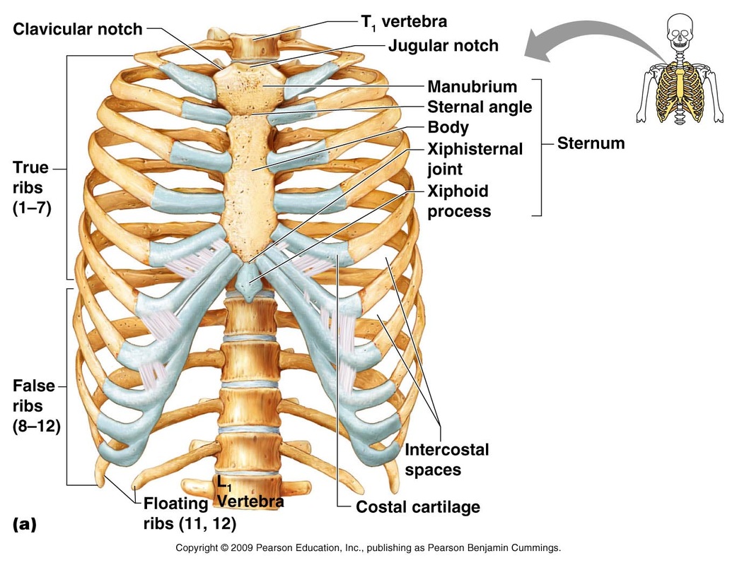
Human rib cage anatomy diagram including anterior and right lateral view all bones surface ... from comps.canstockphoto.com Start studying anatomy of the rib. Vector art, clipart and stock vectors. In this image, you will find thoracic vertebrum, costochondral joint, costal cartilage, costal margin, costal. The shape, size, and structure of body parts permit different fishes to live in different environments or in different parts of the same environment. These are large areas of the cerebral cortex that receive sensory input from multiple different sensory modalities and various association areas and help make associations between various kinds of sensory info. Learn vocabulary, terms and more with flashcards, games and other study tools. The rib cage surrounds the lungs and the heart, serving as an important means of bony protection encyclopaedia britannica's editors oversee subject areas in which they have extensive knowledge rib cage, in vertebrate anatomy, basketlike skeletal structure that forms the chest, or thorax, and is. Rib cage and heart art skeleton print anatomy poster ribs print spine print doctor gift clinic wall decor medicine poster science art.
The serratus anterior muscle originates from a roughened area near the middle of.
Protection on the rib cage of the heart, lungs and diaphragm. Numbered ribs, sternum, cartilage parts and clavicular articulation. Anterior surface of sternum and costal cartilages. Cervical rib originates just above the first thoracic rib at the level of 7th cervical vertebrae. These are large areas of the cerebral cortex that receive sensory input from multiple different sensory modalities and various association areas and help make associations between various kinds of sensory info. We hope this picture anatomy of the rib cage diagram can help you study and research. They articulate with the vertebral column posteriorly, and terminate anteriorly as cartilage (known as costal cartilage). Area between the head and the tubercle of the rib. The shaded areas indicate the extent of the pleural cavities not filled by the lungs. The rib cage is often simplified as an oval shape. Pain+left+side+under+ribs | intro to anatomy 6: This small, rough bump sits on the superointernal border of the horizontally flattened first rib approximately midway between the proximal. As part of the bony thorax, the ribs protect the internal thoracic organs.
Start studying anatomy of the rib. For a gesture drawing, that's good enough. The rib cage is an origin and insertion area for many muscles. The rib cage is often simplified as an oval shape. Posterior, anterior and limbic association areas.
Skeletal System - Life Science from carignanlifescience.weebly.com Diagram of ganglionic areas numbered 1 to 14, used in clinical practice in thoracic oncology for lung cancer disease spread assessment. The shape, size, and structure of body parts permit different fishes to live in different environments or in different parts of the same environment. Rib cage anatomy watercolor this rib cage anatomy art print is a wonderful addition to any interior and will make a perfect v carefully printed to order in our studio original design made by us at codex anatomicus premium heavy blue wall decor rib cage & blue flowers art anatomy diagram | etsy. The rib cage, shaped in a mild cone shape and more flexible than most bone sets, is made up of varying elements such as the thoracic vertebra, 12 equally paired ribs, costal cartilage, and held together anteriorly by the sternum. The ribs, along with the thoracic vertebrae, sternum, and costal cartilages, make up the thoracic cage, also known as the bony thorax. It is responsible for memory, speech, the senses, emotional response, and more. Costal breathing is breathing accomplished by moving of the rib cage as a whole. Related posts of anatomy of ribs and its related area.
These are large areas of the cerebral cortex that receive sensory input from multiple different sensory modalities and various association areas and help make associations between various kinds of sensory info.
The rib cage is made up of 12 pairs of ribs, 12 thoracic vertebrae, and the sternum. The cerebrum is the largest part of the brain. Rib bone anatomy and landmarks. The rib cage, shaped in a mild cone shape and more flexible than most bone sets, is made up of varying elements such as the thoracic vertebra, 12 equally paired ribs, costal cartilage, and held together anteriorly by the sternum. Anatomy is the study of an organism's structures. Different areas often share responsibility for the same task. 12 pairs of anatomical ribs are present in thoracic region — originating from 1st to 12th (t1 to t12) thoracic vertebrae. For a gesture drawing, that's good enough. Instant anatomy is a specialised web site for you to learn all about human anatomy of the body with diagrams, podcasts and revision questions. These are large areas of the cerebral cortex that receive sensory input from multiple different sensory modalities and various association areas and help make associations between various kinds of sensory info. Posterior, anterior and limbic association areas. Numbered ribs, sternum, cartilage parts and clavicular articulation. We have three multimodal association areas:
Diagram of ganglionic areas numbered 1 to 14, used in clinical practice in thoracic oncology for lung cancer disease spread assessment. So, let's learn the ribs so we can attach the muscles in the right place. Human body bones diagram 7 photos of the human body bones diagram 5 major organs of the skeletal system, human body bones diagram quiz, human body skeleton diagram, human skeleton diagram, skeletal map, skeletal structure of the. Learn vocabulary, terms and more with flashcards, games and other study tools. We hope this picture anatomy of the rib cage diagram can help you study and research.
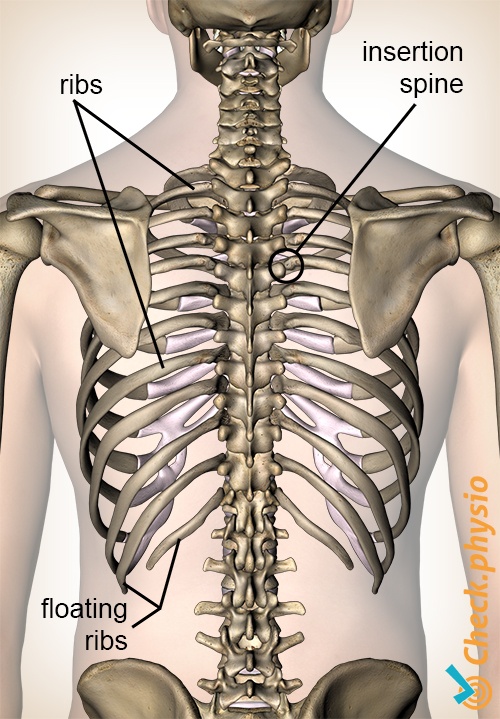
Costochondritis | Physio Check from www.physiocheck.co.uk Start studying anatomy of the rib. Rib cage anatomy watercolor this rib cage anatomy art print is a wonderful addition to any interior and will make a perfect v carefully printed to order in our studio original design made by us at codex anatomicus premium heavy blue wall decor rib cage & blue flowers art anatomy diagram | etsy. 12 pairs of anatomical ribs are present in thoracic region — originating from 1st to 12th (t1 to t12) thoracic vertebrae. Rib cage and heart art skeleton print anatomy poster ribs print spine print doctor gift clinic wall decor medicine poster science art. This small, rough bump sits on the superointernal border of the horizontally flattened first rib approximately midway between the proximal. Human body bones diagram 7 photos of the human body bones diagram 5 major organs of the skeletal system, human body bones diagram quiz, human body skeleton diagram, human skeleton diagram, skeletal map, skeletal structure of the. The ribs are a set of twelve paired bones which form the protective 'cage' of the thorax. Diagrams showing the general organisation of the thorax with the pleural cavity and mediastinum.
For a gesture drawing, that's good enough.
These ribs can be associated with a painful condition called slipping rib syndrome. For more anatomy content please follow us and visit our website: Different areas often share responsibility for the same task. Vector art, clipart and stock vectors. Find this pin and more on anatomy and science of the human body by jordan cobb. The rib cage is an origin and insertion area for many muscles. We have three multimodal association areas: The rib cage, shaped in a mild cone shape and more flexible than most bone sets, is made up of varying elements such as the thoracic vertebra, 12 equally paired ribs, costal cartilage, and held together anteriorly by the sternum. They articulate with the vertebral column posteriorly, and terminate anteriorly as cartilage (known as costal cartilage). Pain+left+side+under+ribs | intro to anatomy 6: Costal breathing is breathing accomplished by moving of the rib cage as a whole. The shape, size, and structure of body parts permit different fishes to live in different environments or in different parts of the same environment. Posterior, anterior and limbic association areas.

0 Comments:
Posting Komentar