Knee Muscle Anatomy Mri - Mri Knee Anatomy Knee Sagittal Anatomy Free Cross Sectional Anatomy : Tips to keep joints healthy.. General anatomy and musculoskeletal system. Knee anatomy francesc malagelada jordi vega pau golanó the knee is the largest joint in the human body and one of the most complex from a functional point of view. Learn about the muscles, tendons, bones, and ligaments that comprise the knee joint anatomy. Click now to learn more about the bones, muscles, and soft tissues of these regions at leg and knee anatomy: This section of the website will explain large and minute details of sagittal knee use the mouse scroll wheel to move the images up and down alternatively use the tiny arrows (>>) on both side of the image to move the images.
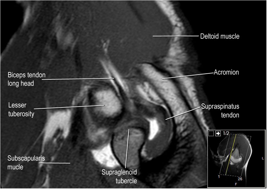
Knowing about knee anatomy can help people understand how knee arthritis develops and sometimes causes pain. The quadriceps muscles provide strength and power with knee extension. Normal knee mri for reference. Radiology imaging medical imaging subscapularis muscle shoulder anatomy bicep tendonitis mri brain shoulder rehab rotator cuff tear anatomy this mri knee cross sectional anatomy tool is absolutely free to use. Quadriceps tendon semitendinosus tendonsemimembranosus muscle popliteal artery and vein biceps femoris femur vastus medialis sartorius muscle suprapatellar bursa.
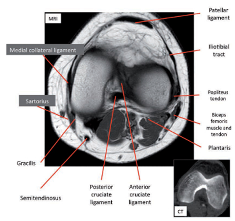
How To Read The Normal Knee Mri Kenhub from thumbor.kenhub.com Rubin da, kettering jm, towers jd, britton ca: Magnetic resonance imaging (mri) is the test of choice to confirm the diagnosis of a torn meniscus. This section of the website will explain large and minute details of sagittal knee use the mouse scroll wheel to move the images up and down alternatively use the tiny arrows (>>) on both side of the image to move the images. Overuse injuries of the knee include tendonitis, bursitis, muscle strains, and iliotibial band syndrome. Each anatomical structure was labeled interactively. The knee is designed to fulfill a number of functions: Magnetic resonance imaging (mri scan): This webpage provides a gallery of images that presents the anatomical structures found on knee mri.
Please email baodo at stanford.edu.
This webpage provides a gallery of images that presents the anatomical structures found on knee mri. Scroll through the structures to understand the anatomy. Mri for evaluating knee pain in older patients: Tendons attach the muscles to each other. The knee joint is the junction of the thigh and leg. Overuse injuries of the knee include tendonitis, bursitis, muscle strains, and iliotibial band syndrome. Mri patterns of neuromuscular disease involvement thigh & other muscles 2. Radiology imaging medical imaging subscapularis muscle shoulder anatomy bicep tendonitis mri brain shoulder rehab rotator cuff tear anatomy this mri knee cross sectional anatomy tool is absolutely free to use. Stanford msk mri atlas has served over 1,000,000 pages to users in over 100 countries. The knee joint is most significantly affected by two major muscle groups: Normal knee mri for reference. Free cross sectional anatomy of the knee based on mri : This mri knee cross sectional anatomy tool is absolutely free to use.
The muscles that affect the knee's movement run along the thigh and calf. Involved early gray = muscle: Click now to learn more about the bones, muscles, and soft tissues of these regions at leg and knee anatomy: View of the anatomical labels. Normal knee mri for reference.
The Radiology Assistant Shoulder Anatomy Mri from radiologyassistant.nl Click on the links to show each structure. Magnetic resonance imaging (mri scan): Radiology imaging medical imaging subscapularis muscle shoulder anatomy bicep tendonitis mri brain shoulder rehab rotator cuff tear anatomy this mri knee cross sectional anatomy tool is absolutely free to use. Use the checklist to quiz yourself. This mri knee cross sectional anatomy tool is absolutely free to use. The knee is designed to fulfill a number of functions: Each anatomical structure was labeled interactively. Mri for evaluating knee pain in older patients:
Support the body in an upright position without the need for muscles to work.
If you think of the knee in layers, the deepest layer is bone and ligaments, then ligaments of the joint capsule, then muscles on top. Tips to keep joints healthy. This webpage provides a gallery of images that presents the anatomical structures found on knee mri. Please email baodo at stanford.edu. Magnetic resonance imaging (mri) is the test of choice to confirm the diagnosis of a torn meniscus. Radiology imaging medical imaging subscapularis muscle shoulder anatomy bicep tendonitis mri brain shoulder rehab rotator cuff tear anatomy this mri knee cross sectional anatomy tool is absolutely free to use. Learn about the muscles, tendons, bones, and ligaments that comprise the knee joint anatomy. Click on the links to show each structure. Each anatomical structure was labeled interactively. Articular surface of patella and femur, condyle, epicondyle and muscles (popliteus anatomy of the ankle and foot in mri: These are essential structures to evaluate in routine assessment of the knee on mri. Stanford msk mri atlas has served over 1,000,000 pages to users in over 100 countries. Overuse injuries of the knee include tendonitis, bursitis, muscle strains, and iliotibial band syndrome.
Involved early gray = muscle: On anatomical parts the user. Tips to keep joints healthy. Articular surface of patella and femur, condyle, epicondyle and muscles (popliteus anatomy of the ankle and foot in mri: Knowing about knee anatomy can help people understand how knee arthritis develops and sometimes causes pain.
Knee Springerlink from media.springernature.com The quadriceps muscles provide strength and power with knee extension. The muscles of the knee include the quadriceps, hamstrings, and the muscles of the calf. Knee anatomy francesc malagelada jordi vega pau golanó the knee is the largest joint in the human body and one of the most complex from a functional point of view. Please email baodo at stanford.edu. Learn about the muscles, tendons, bones, and ligaments that comprise the knee joint anatomy. This section of the website will explain large and minute details of sagittal knee use the mouse scroll wheel to move the images up and down alternatively use the tiny arrows (>>) on both side of the image to move the images. Involved early gray = muscle: Quadriceps tendon semitendinosus tendonsemimembranosus muscle popliteal artery and vein biceps femoris femur vastus medialis sartorius muscle suprapatellar bursa.
Learn about mri anatomy with free interactive flashcards.
The knee joint is most significantly affected by two major muscle groups: These are essential structures to evaluate in routine assessment of the knee on mri. Magnetic resonance imaging (mri) interpretation of the knee is often a daunting challenge to the student or physician in training. Mr imaging of knees having isolated and combined ligament injuries. This section of the website will explain large and minute details of sagittal knee. Each anatomical structure was labeled interactively. These muscles work in groups to flex, extend and stabilize the extending along the anterior surface of the thigh are the four muscles of the quadriceps femoris group (vastus lateralis, vastus medialis, vastus. Use the checklist to quiz yourself. Learn about the muscles, tendons, bones, and ligaments that comprise the knee joint anatomy. Want to learn more about it? Muscle anatomy lats 12 photos of the muscle anatomy lats back muscles anatomy lats, muscle anatomy lats, human muscles, back muscles anatomy lats, muscle anatomy lats. Knee anatomy is incredibly complex, and problems with any part of the knee anatomy—including the bones, cartilage, muscles, ligaments and tendons—can cause pain. The journal of musculoskeletal medicine.
0 Comments:
Posting Komentar